| dc.contributor.author | Mande, J. O. | |
| dc.contributor.author | Mbithi, Peter Mulwa F | |
| dc.contributor.author | Mbugua, S.W. | |
| dc.contributor.author | Buoro, I. B. J. | |
| dc.contributor.author | Gathumbi, P.K. | |
| dc.date.accessioned | 2013-02-22T09:12:23Z | |
| dc.date.issued | 2003 | |
| dc.identifier.citation | Journal of South African Veterinary Association (2003)74(1): 11-13 | en |
| dc.identifier.uri | http://erepository.uonbi.ac.ke:8080/xmlui/handle/123456789/10723 | |
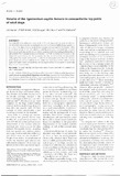
| dc.description.abstract | Ventrodorsal pelvic radiographs were made of 32 adult dogs under general anaesthesia. The hip joints were evaluated according to the severity of osteoarthritic changes graded as 0,1,2 or 3. The dogs were euthanased, the hip joints opened and the lignlllelltulll capitis fellloris dissected out ill toto. The volume of each ligament was determined using a water displacement technique and the mean volume compared to the four radiographic grades of osteoarthritis. There was an inverse correlation (r = -0.75) between the mean volume of the ligamentum capitis femoris and the increasing severity of osteoarthritis as assessed by radiography. The results confirmed the crucial role of radiography in the clinical evaluation of hip dysplasia and osteoarthritis in the adult dog. Assessment of the volume of the ligamentum capitis femoris revealed that it is an important tool for research in canine hip dysplasia and osteoarthritis. | en |
| dc.language.iso | en | en |
| dc.subject | Hip dysplasia | en |
| dc.subject | Hip joint | en |
| dc.subject | Ligamentum capitis femoris | en |
| dc.subject | Osteoarthritis | en |
| dc.subject | Ventrodorsal pelvic radiography | en |
| dc.title | Volume of the ligamentum capitis femoris in osteoarthritic hip joints of adult dogs | en |
| dc.type | Article | en |
| local.publisher | Department of clinical studies | en |