| dc.contributor.author | Onditi, Faith I | |
| dc.contributor.author | Nyamongo, Onkoba W | |
| dc.contributor.author | Omwandho, Charles O | |
| dc.contributor.author | Maina, Naomi W | |
| dc.contributor.author | Maloba, Fredrick | |
| dc.contributor.author | Farah, Idle O | |
| dc.contributor.author | King, Christopher L | |
| dc.contributor.author | Moore, Julie M | |
| dc.contributor.author | Ozwara, Hastings S | |
| dc.date.accessioned | 2015-08-07T15:17:36Z | |
| dc.date.available | 2015-08-07T15:17:36Z | |
| dc.date.issued | 2015-03-18 | |
| dc.identifier.citation | Malaria Journal. 2015 Mar 18;14(1):118 | |
| dc.identifier.uri | http://dx.doi.org/10.1186/s12936-015-0631-5 | |
| dc.identifier.uri | http://hdl.handle.net/11295/89655 | |
| dc.description.abstract | Abstract
Background
Placental malaria (PM) causes adverse pregnancy outcomes in the mother and her foetus. It is difficult to study PM directly in humans due to ethical challenges. This study set out to bridge this gap by determining the outcome of PM in non-immune baboons in order to develop a non-human primate model for the disease.
Methods
Ten pregnant baboons were acquired late in their third trimester (day 150) and randomly grouped as seven infected and three non-infected. Another group of four nulligravidae (non-pregnant) infected was also included in the analysis of clinical outcome. Malaria infection was intravenously initiated by Plasmodium knowlesi blood-stage parasites through the femoral vein on 160th day of gestation (for pregnant baboons). Peripheral smear, placental smear, haematological samples, and histological samples were collected during the study period. Median values of clinical and haematological changes were analysed using Kruskal-Wallis and Dunn’s Multiple Comparison Test. Parasitaemia profiles were analysed using Mann Whitney U test. A Spearman’s rank correlation was run to determine the relationship between the different variables of severity scores. Probability values of P <0.05 were considered significant.
Results
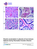
Levels of white blood cells increased significantly in pregnant infected (34%) than in nulligravidae infected baboons (8%). Placental parasitaemia levels was on average 19-fold higher than peripheral parasitaemia in the same animal. Infiltration of parasitized erythrocytes and inflammatory cells were also observed in baboon placenta. Malaria parasite score increased with increase in total placental damage score (rs = 0.7650, P <0.05) and inflammatory score (rs = 0.8590, P <0.05). Although the sample size was small, absence of parasitized erythrocytes in cord blood and foetal placental region suggested lack of congenital malaria in non-immune baboons.
Conclusion
This study has demonstrated accumulation of parasitized red blood cells and infiltration of inflammatory cells in the placental intravillous space (IVS) of baboons that are non-immune to malaria. This is a key feature of placental falciparum malaria in humans. This presents the baboon as a new model for the characterization of malaria during pregnancy. | |
| dc.title | Parasite accumulation in placenta of non-immune baboons during Plasmodium knowlesi infection | |
| dc.type | Journal Article | |
| dc.date.updated | 2015-08-07T15:17:36Z | |
| dc.language.rfc3066 | en | |
| dc.rights.holder | Onditi et al.; licensee BioMed Central. | |